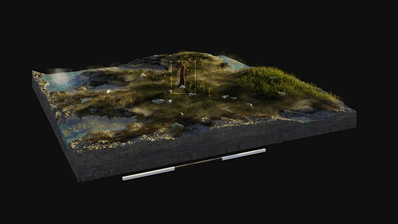
Prototaxites — Illustration by Matthew Humpage
Prototaxites was an extinct lineage of multicellular terrestrial eukaryotes. Prototaxites was the first giant organism to live on the terrestrial surface, reaching sizes of 8 metres in the Early Devonian.
However, its taxonomic assignment has been debated for over 165 years. Tentative assignments to groups of multicellular algae or land plants have been repeatedly ruled out based on anatomy and chemistry resulting in two major alternatives: Prototaxites was either a fungus or a now entirely extinct lineage.
Recent studies have converged on a fungal affinity. Here we test this by contrasting the anatomy and molecular composition of Prototaxites with contemporary fungi from the 407-million-year-old Rhynie chert.
We report that Prototaxites taiti was the largest organism in the Rhynie ecosystem and its anatomy was fundamentally distinct from all known extant or extinct fungi. Furthermore, our molecular composition analysis indicates that cell walls of P. taiti include aliphatic, aromatic, and phenolic components most similar to fossilisation products of lignin, but no fossilisation products characteristic of chitin or chitosan, which are diagnostic of all groups of extant and extinct fungi, including those preserved in the Rhynie chert.
We therefore conclude that Prototaxites was not a fungus, and instead propose it is best assigned to a now entirely extinct terrestrial lineage.

Prototaxites taiti material from the Rhynie chert. a-c, Images of two of the four thin sections containing the fragments that constitute the P. taiti type material, including medullary spots (b) and peripheral region (c). d-l: P. taiti material used in this study. d,e: d, Lyon 156 with P. taiti highlighted in dashed lines. e, thin section produced from the block in (d) showing the fractured P. taiti specimen. f, Magnified image of the thin section in (e) showing the characteristic tubes and medullary spots of P. taiti. g, h: g, thin section made from Lyon 48 with P. taiti in dashed box. h, detail of thin section in (g) showing the tubes. i m: Imaging and reconstruction of a large, exceptionally well-preserved P. taiti from NSC.36. i, Photogrammetry model of NSC.36 before cutting with surface exposed P. taiti circled by dashed line. j, Photogrammetry model of NSC.36 after initial cutting of the block with P. taiti circled by a dashed line. k, Block of NSC.36 from which thin sections were produced, showing medullary spots throughout the body. l, thin section taken from the block in (k) showing characteristic tubes and medullary spots of P. taiti. m, Artist reconstruction of P. taiti within the Rhynie ecosystem including hypothesised reconstruction of the aerial portion. Illustration by Matthew Humpage, Northern Rouge Studios. Scale bars: 3m (m), 3 cm (i), 2cm (j), 1cm (d), 5mm (e, g, k), 1mm (c), 500µm (a, l), 200µm (b, f), 100µm (h). Specimen accession codes: GLAHM Kid 2523 (a, b), GLAHM Kid 2525 (c), Lyon 156 (d), Lyon 156 MPEG0078 (e, f), NSC.36 (i-k), NMS G.2024.5.7 (l). — biorxiv.org
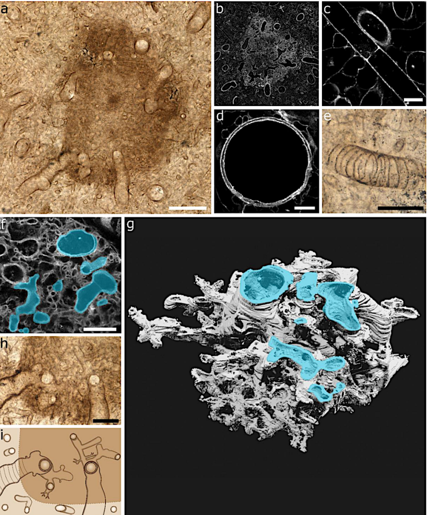
The medullary spots and tube types of Prototaxites taiti are morphologically distinct from anything observed in extinct or extant fungal groups, and do not support a fungal affinity for Prototaxites. a, Transmitted light image showing a medullary spot within the body of P. taiti. b, The same medullary spot imaged using CLSM, showing the spot to be composed of densely packed fine tubes contrasting with the less densely packed body. c-e, Details of tubes types 1-3 seen in the body of P. taiti: small diameter type 1 tube with septal pore (c), larger diameter type 2 tube (d), and type 3 tube with annular thickenings (e). f-h, Airyscan CLSM three-dimensional imaging reveals that at the medullary spot region all tube types are connected through a highly branched network. Tubes of a variety of morphologies (highlighted in cyan in f and g) were found to be connected to each other in a dense and fine made available under aCC-BY-NC-ND 4.0 International license. (which was not certified by peer review) is the author/funder, who has granted bioRxiv a license to display the preprint in perpetuity. It is bioRxiv preprint doi: https://doi.org/10.1101/2025.03.14.643340; this version posted March 17, 2025. The copyright holder for this preprint branching network through the construction of a 3D SPIERS model (g) using Airyscan CLSM z stack data (the first image in the stack is shown in f). Examination of the spot region (h) supports the interconnection of all tube types through fine branching at the medullary spots, as shown in the schematic in (i). Scale bars: 100µm (a), 50µm (e, h), 20 µm (f), 10µm (c,d). Specimen accession code: NMS G.2024.5.7. — biorxiv.org
Prototaxites was an extinct lineage of multicellular terrestrial eukaryotes, biorxiv.org (open access)
Astrobiology